
Giant cell tumour:
Definition: It is an osteolytic of uncertain origin appears mostly in epiphysis in the long bone.
Other names:
- Osteoclastoma
- Giant cell sarcoma
- Benign giant cell tumor
Because of it’s rarely metastatic character giant cell tumor is a locally malignant tumor because rarely metastasis.
Age of onset: 20-45 years (most common in female, usually 3rd decades of life).
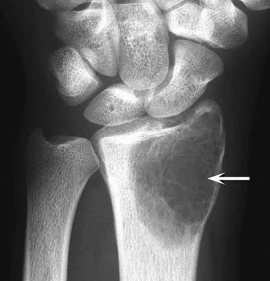
Site of onset: Around knee, proximal humerus, distal radius (most common).
History of trauma present (may be)
Chief complaint:
1.Pain
2. Swelling –
👉Towards one side
👉 Skin over the lesion is shiny
👉 Egg shell cracking sound may be on applying pressure.
At the beginning there’s no changes in joint movement but later joint movement diminished.
Investigation:
- X-ray
- CT scan
- MRI (Magnetic resonance imaging)
- Arthroscopy

Biochemical test:
- Serum Calcium
- Serum phosphate
- Serum alkaline phosphatase
- Routine blood test
X-ray findings :
- Round/oval osteolytic lesion (epiphyseal, eccentric, expanded size).
- The cortex is thin & ballooned.
Treatment :
👉 Generally treatment depends on size of tissue bone involvement, staging & grading.
- Curettage only (85% recurrent)
- Bone curettage with bone graft (40%).
- Chemical cauterization + curettage
- Curettage + cauterization + bone graft
- Curettage + chemical cauterization + Bone Cement
- Curettage + cryosurgery with liquid nitrogen + bone graft.
- Margin excision + bone graft (36%)
- Wide excision + bone graft
- Amputation
If pulmonary metastasis occur-
- Embolization
- Excision
Sazzad mahmud
Monowara Sikder Medical College (2015-16).
Platform Academic / Gazi Abdullah Al Mamun