

Placenta এর Trophoblast এর অস্বাভাবিক growth এর কারণে কিছু conditions পাওয়া যায় যাদের আমরা Gestational trophoblastic disease (GTD) বলি। এদের মধ্যে একটি condition হচ্ছে Hydatidiform mole।
আমরা আজকে আলোচনা করব Hydatidiform mole নিয়ে:
এখানে Placenta এর young chorionic villi তে কিছু degenerative changes, আবার কিছু Proliferative changes পাওয়া যায়।
এটা একটা Benign neoplasia with malignant potential।
একে আমরা দুইভাগে ভাগ করে থাকি:
a) Complete mole
b) Partial mole
কেন হচ্ছে?
-Due to abnormal fertilization.
Complete mole এর ক্ষেত্রে যে ovum টা থাকে সেটায় nucleus থাকেনা। এটা সম্পূর্ণ Paternal origin।
Karyotype হবে: 46,XX অথবা 46,XY।
এই ক্ষেত্রে Embryo একদমই grow করতে পারে না। এখানে শুধুই Placenta থাকে, কোনো বাচ্চা থাকেনা।
যত Chorionic villi থাকে সবগুলোর hydropic change হয়ে যায়। তাই এর নাম Complete mole।
Partial mole এর ক্ষেত্রে ovum ঠিক থাকছে কিন্তু embryo তে ক্রোমোজোম একসেট বেশী থাকে। (মায়ের একসেট + বাবার দুইসেট ক্রোমোজোম)।
Karyotype: Triploid, 69,XXX or 69,XXY (90%).
Normal villi এর সাথে Hydropic villi পাওয়া যাবে, fetus ও পাওয়া যাবে।
তাই এর নাম Partial mole।

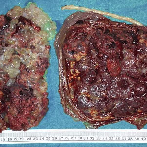
Pathology of Hydatidiform mole-
এখানে,
Trophoblastic cells এর hyperplasia হয়।
⬇️
সেখান থেকে যে Secretion আসে তা maternal blood এর materials এর সাথে মিলে villous গুলোতে জমা হয়ে hydropic change করে, আর vesicle তৈরী করে (যার কারণে একে Vesicular mole ও বলা হয়)।
এই ফ্লুইড যেহেতু Interstitial fluid, তাই এর composition ও ascitic/edema fluid এর মতোই, পার্থক্য শুধু এই যে – এখানে Human chorionic gonadotropin উপস্থিত।
Microscope এ চোখ রাখলে আমরা দেখতে পাই,
- Stromal tissue তে hydropic degeneration এর কারণে তা সংকুচিত হয়ে আছে।
- Cytotrophoblast & sycytiotrophoblast এর দিকে তাকালে marked proliferation দেখা যাবে।
- Villi তে blood vessels অনুপস্থিত।
খালি চোখে দেখলে:
- অসংখ্য cyst এর cluster হিসেবে দেখা যাবে।
- এইখানে যেহেতু Beta hCG excessive থাকে, তাই এর প্রভাবে Ovary তে আমরা Theca lutein cyst পেতে পারি।
Hydatidiform mole এর চিকিৎসা করলে দুইমাস পর তা spontaneously কমে যায়।
Management of Hydatidiform mole-
Diagnosis এর জন্য যখন History নিব:
- Patient আমাদের কাছে এসে short period of amenorrhoea (৮-১২ সপ্তাহ) এর কথা বলবে।
- Irregular heavy vaginal bleeding এর কথা বলবে (এটা কমন প্রেজেন্টেশন, ৯০%)।
- এই Blood এর সাথে gelatinous fluid থাকতে পারে যেটাকে বলা হয় ‘White current in red current juice’।
- Vagina দিয়ে Grape like vesicle বের হওয়ার কথা বলবে (diagnostic of vesicular mole)।
- Lower abdominal pain থাকতে পারে-
- Uterus এর overstreching জন্য,
- Concealed hemorrhage এর জন্য,
- Invasive mole যদি uterus কে perforation করে সেই কারণে,
- Infection হলে,
- Uterus তার ভিতরের জিনিসগুলো বের করার জন্য যদি contraction করে সেই কারণেও ব্যাথা হতে পারে,
-(HCG বেড়ে যাওয়ার কারণে) Hyperemesis এর complaint করতে পারে।
General examination করলে-
- Patient is Ill looking,
- Pallor পাওয়া যাবে, out of proportion to blood loss (concealed hemorrhage হয় তাই)।
- Features of pre-eclampsia পেতে পারি।
Abdominal examination করলে-
- Uterus এর size is more than the period of amenorrhoea। কেন? – ওই যে বললাম Villus গুলো বড় হয়ে যাচ্ছে, Concealed haemorrhage ও থাকতে পারে।
- Firm, elastic, doughy feel of uterus. কেন?- এখানে তো কোনো Amniotic fluid sac ই নেই তাই।
- বাচ্চা নেই তাই কোনো Fetal part, Heart sound ও পাচ্ছি না।
Vaginal examination করলে-
- Vaginal discharge এ vesicles পাওয়া যাবে(pathognomonic)।
- Fornix এ হাত দিলে bilateral enlargement of ovary পেতে পারি (Theca lutein cyst এর ক্ষেত্রে)।
- Cervix এর os যদি open থাকে সেক্ষেত্রে placental membrane এর বদলে হাতে আসবে blood clot, vesicles।
Investigation :
a) Blood grouping, Rh typing, CBC,
b) Ultrasonography of the uterus,
c) adnexa-
*এখানে Snow storm appearance of the mole পাওয়া যাবে, এটাই diagnostic feature।
*Serum hCG markedly elevated থাকবে।
d) Chest X-ray করতে হবে কোনো pulmonary embolization এর evidence আছে কি না দেখার জন্য। Metastatic deposit যদি থাকে তাহলে:
a) Cannon ball opacity’ দেখা যাবে।
b) Liver, renal & thyroid function test গুলোও করে নিতে পারি।
Treatment of Hydatidiform mole:
Patient যেহেতু bleeding নিয়েই commonly present করে তাই তাকে supportive treatment দেওয়া লাগতে পারে।
- I/V fluid দিব
- Pallor বেশী থাকলে blood transfusion করব
- Parenteral antibiotics দিব যদি infection থাকে।
Definitive treatment:

- Suction evacuation & curettage is preferred.
মনে রাখতে হবে, এক্ষেত্রে আগেই Oxytocin দিলে expulsion সহজ হয় কিন্তু pulmonary embolization এর chances বেড়ে যায় তাই evacuation এর পরে methergine 0.2 mg IM দেয়া হয় যেনো bleeding কমে। - যাদের বয়স >40 years & Family complete
অথবা, evacuation এর সময় যদি uterus perforated হয়ে যায়/uncontrolled bleeding হয় তাদের Hysterectomy is the choice of treatment। - Evacuation এর পরে Rh (-ve) nonimmunized patient কে Anti D immunoglobulin দিতে হবে।
Faria
Session :14-15
JRRMC
প্ল্যাটফর্ম একাডেমিক/ তাবাসসুম ইসলাম