
Leukaemia আসলে কি?🙄
🚩Leukaemia একটা cancerous condition যেটা blood forming tissue (mainly bone marrow) থেকে arise করে।
Normally blood cell গুলো bone marrow থেকে তৈরী হয়। Bone marrow এর stem cell কে 2 type এ ভাগ করা যায়–
1) Myeloid stem cell.
2) Lymphoid stem cell.
Myeloid stem cell থেকে mature blood cell (RBC, WBC, Platelets) arise করে।
Lymphoid stem cell থেকে প্রথমে Lymphoblast & then B lymphocyte, T lymphocyte ও Natural killer cell arise করে (এরা body এর immune system কে maintain করে)।
🚩Leukaemia mainly granulocytic & lymphoid series কে affect করে, erythroid & megakaryocytic series কে rarely affect করে।
That’s why, leukaemia patient দের WBC, lymphoblast & lymphocyte count বেশি থাকে এবং RBC, Platelet count কম থাকে।
💬Leukaemia তে haemopoietic stem cell গুলোর abnormal proliferation হবে। তারপর abnormally proliferated cell গুলো প্রথমে bone marrow তে জমা হতে থাকবে এবং পরবর্তীতে liver, spleen, lymph node & other lymphatic tissue তে জমা হবে। Abnormal cell গুলো জমা হতে হতে একটা neoplastic condition arise করবে।So, by definition we can say–
💁Leukaemia is a neoplastic disorder characterized by abnormal proliferation of haemopoietic stem cell causes progressively increasing infiltration of bone marrow & in most cases liver, spleen, lymph nodes & other lymphatic tissue.
🚩এখন আসি Leukaemia কি কি কারণে হয়?🤔
🔎Causes of Leukaemia :
1) Chromosomal abnormalities.
2) Radiation:
★ X- ray,
★ Radio therapy,
★Children who are exposed to diagnostic X-ray in utero.
3) Chemical agent:
★Chronic inhalation of benzene,
★Cytotoxic drugs:
Melphalan, Procarbazine, Chlorambucil.
4) Virus : Retrovirus.
5) Immunological deficiency state : Hypogammaglobulinaemia.
🚩এবার Leukaemia কে classify করা যাক–
💁According to clinical course & predominant cell involvement :-
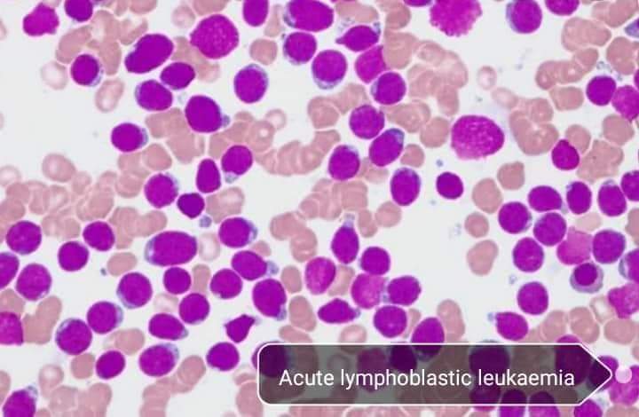
1) Acute lymphoblastic leukaemia (ALL)
2) Acute myeloid leukaemia (AML)
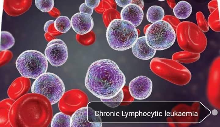
3) Chronic lymphocytic leukaemia (CLL)
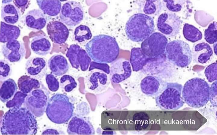
4) Chronic myeloid leukaemia ( CML)
👉এছাড়া French- American -British haematologist রা morphology এর উপর depend করে leukaemia কে classify করেছেন। কিন্তু এখন এটি ব্যবহার করা হয় না।
FAB (French- American- British) classification :-
A) Acute leukaemia:
1)Acute myeloblastic leukaemia (AML):
M0- Minimally differentiated AML
M1- Myeloblastic leukaemia without maturation.
M2- Myeloblastic leukaemia with maturation.
M3- Hypergranular promyelocytic leukaemia.
M4- Myelomonocytic leukaemia.
M5- Monocytic leukaemia.
M6- Erythro leukaemia.
M7- Megakaryoblastic leukaemia.
2) Acute lymphoblastic leukaemia (ALL):
L1- Small homogenous lymphoblast.
L2- Large heterogenous lymphoblast.
L3- Large homogenous lymphoblast.
B) Chronic leukaemia:
1) Chronic myeloid leukaemia.
2) Chronic lymphocytic leukaemia.
🚩Clinical presentation of leukaemia:
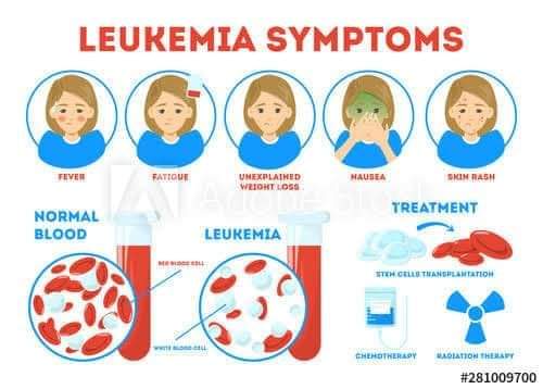
🔎Symptoms:
🔸Anaemia,
🔸 Leucopenia(WBC count is exceed but it has qualitative defect),
🔸Thrombocytopenia,
🔸Marrow infiltration,
🔸Weight loss,
🔸Abdominal pain,swelling &
discomfort.
🔎Sign:
🔹Pallor due to anaemia,
🔹Fever followed by recurrent infection
due to leukopenia,
🔹Petechiae purpura, bruise due to
thrombocytopenia,
🔹Bone pain, bony tenderness due to
marrow infiltration,
🔹Lymphadenopathy,
🔹 Hepatosplenomegaly.
🙂এবার আসি acute leukaemia তে-
🚩এখানে immature blast cell গুলো অর্থাৎ myeloblast, lymphoblast গুলোর uncontrol proliferation হবে এবং malignant condition progress করবে।
👉Basically acute leukaemia কে 2 type এ ভাগ করা যায়–
1) Acute myeloblastic leukaemia (AML).
2) Acute lymphoblastic leukaemia (ALL)
ALL তে usually children (1-5 years) রা affected হয় এবং AML তে adult রা affected হয়।
ALL এর ক্ষেত্রে lymph node enlarge হয়ে যায় কিন্তু ALL এর prognosis ভাল। আবার AML এর prognosis খারাপ।
★Clinically AML & ALL কে কিভাবে differentiate করতে পারি?? 🤔
⏩Lab diagnosis:
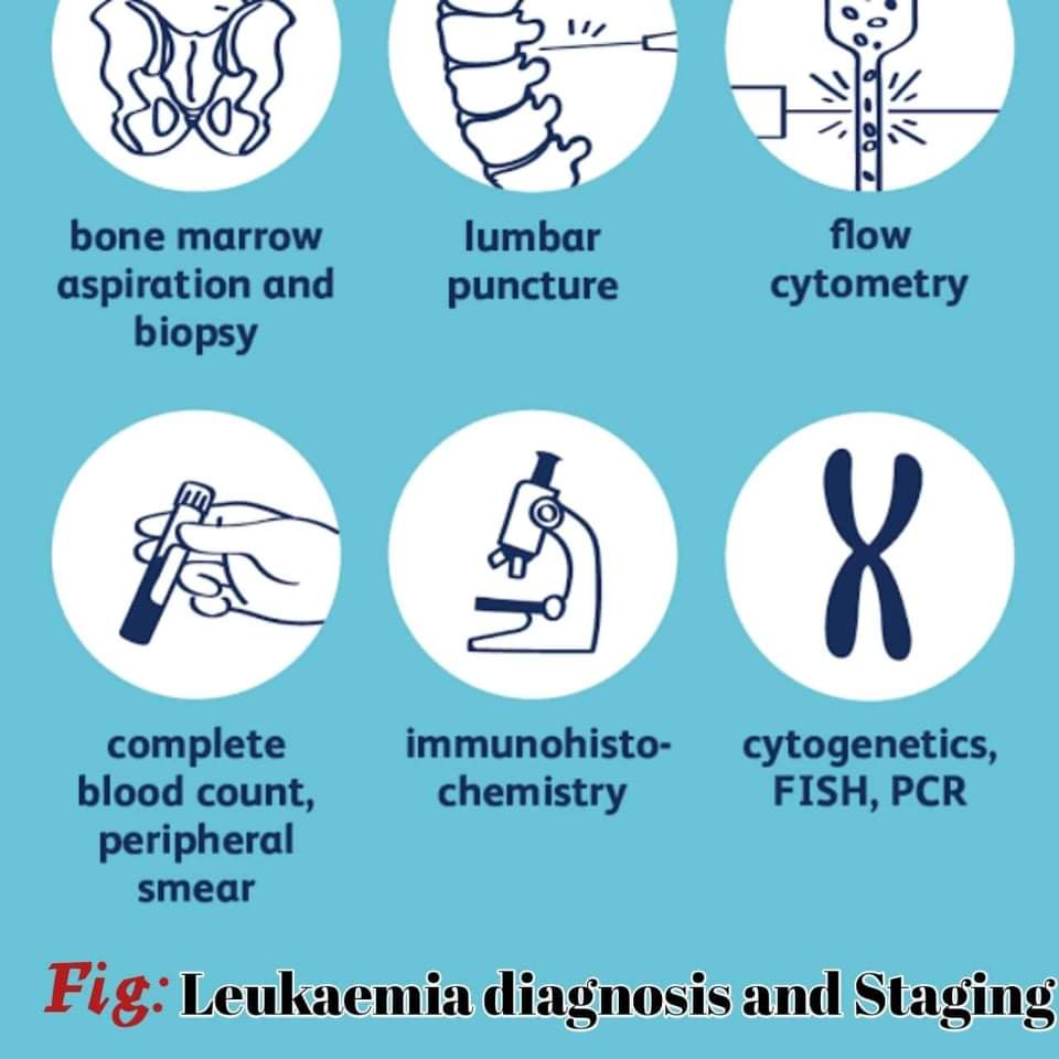
🚩Blood picture দেখব এবং bone marrow examination করতে পারি।
🚩Blood picture এ 🔺Hb%
🔺PCV
🔺Peripheral blood film দেখব।
AML & ALL এর ক্ষেত্রে difference ঘটে শুধুমাত্র WBC তে; বাকী সব same থাকে।
1) Hb% : Reduced in both AML & ALL যেহেতু leukaemia erythroid series কে affect করে না।
2) PCV: As RBC count reduced, so PCV is also reduced.
3) Peripheral blood film:
🌀RBC: RBC count decrease করে, but RBC size normocytic & normochromic থাকে। Sometimes it may be anisocytosis ( crescent shape) এবং poikilocytosis (biconcave shape টা flat হয়ে যায়)।
🌀WBC: যেহেতু leukaemia myeloid series কে affect করে, তাই WBC count increased হয়।💧The count is : 20,000-1,00,000/mm3 of blood.
💧Immature blast cell increase (80-90%).
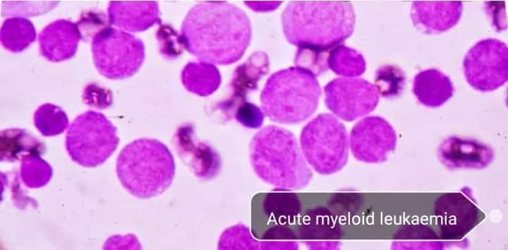
💧AML এ blast cells are myeloblast, monoblast.
💧ALL এ blast cells are lymphoblast.
⚫Myeloblast, lymphoblast এর থেকে দেখতে একটু বড়।
💧Myeloblast এ auer body present থাকে but lymphoblast এ auer body থাকে না।
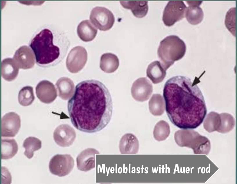
Auer body একটা rod shaped structure যেটা myeloid, myelocyte & monocyte এর cytoplasm এ present থাকে।
💧ALL তে smear cell present থাকে but AML তে smear cell absent.
💧Myeloblast এ 3-4 টা nucleoli থাকে & chromatin network loose থাকে।
Lymphoblast এ 0-2 টা nucleoli থাকে & chromatin network dense.
💧Myeloblast এর cytoplasm relatively more than lymphoblast. এজন্য nucleus/cytoplasm ratio myeloblast এ কম এবং lymphoblast এর ক্ষেত্রে বেশি হয়।
🌀 Platelet: Leukaemia megakaryocyte series কে affect করে না, so platelet count decrease এবং Thrombocytopenia occurs both in AML & ALL.
Reticulocyte: Upto 5% in AML & ALL.🚩এবার আসি bone marrow examination:
এখানে AML & ALL এর same result পাওয়া যায়।
Cellularity: যেহেতু WBC count increase করে, so
cellularity is hypercellular.
M/E ratio: WBC count increase করার কারণে
ratio increased হয়।
Erythropoiesis: Reduced.
Leukopoiesis: Increased.
megakaryopoiesis: Reduced.
🙂 Chronic leukaemia:
🚩Acute leukaemia rapidly progress করে but chronic leukaemia slowly progress করে। Chronic leukaemia এর ক্ষেত্রে well differentiated leukocyte এর uncontrolled proliferation হয় এবং এর prognosis খারাপ।
👉Chronic leukaemia ও 2 type:
1) Chronic myeloid leukaemia (CML).
2) Chronic lymphocytic leukaemia (CLL).
★Clinically এদের কিভাবে differentiate করতে পারি?🙄
🚩এখানেও আমরা blood picture দেখব & bone marrow examination করব।
🚩Blood picture এ 🔻Hb%
🔻Peripheral blood film দেখব।
1) Hb% : Reduced both in CML & CLL.
2) Peripheral blood film :
🌀 RBC: যেহেতু leukaemia erythroid series কে affect করে না, তাই RBC are normocytic & normochromic.
Sometimes basophilic stippling present in CML.
Sometimes normocytic & normochromic with spherocytosis.
🌀WBC :
🔴In CML:
–WBC count is >50×10^9/L
— Myeloid cell present.
— Basophil count increased.
🔴In CLL:
–WBC count is 50,000-2,00,000/mm3
— Lymphocyte count- total WBC এর 90%
— Smudge cell/busket cell present ( the cell that does not have cytoplasmic membrane or nuclear structure).
🌀 Platelet: In all type of leukaemia, platelet count decreased & Thrombocytopenia occurs except chronic phase of CML. 👉In chronic phase of CML > platelet count increase.
But later stage > platelet count decrease.
👉
🌀Reticulocyte: Count is moderate in CML but reticulocytosis occurs in CLL.
🚩এবার Bone marrow examination :-
Cellularity: WBC count বেশি থাকার জন্য hypercellular হয়।
M/E ratio: As WBC count increased, so ratio is increased.
Erythropoiesis: Decreased.
Leukopoiesis: Increased.
💧 In CML- Immature cells are myelocyte & myeloblast.
💧 In CLL- Immature cells are lymphocyte.
Megakaryopoiesis: Decreased.
🚩🚩The main difference between CML & CLL is chromosomal abnormality.
🙂>> presence of Philadelphia chromosome is the diagnostic feature of CML. এটা 90% CML patient দের present থাকে।
এখানে,
chromosome 22 & 9 এর মধ্যে reciprocal translocation হয়।chromosome 9 এর C- abl part, chromosome 22 এর BCR part এ transfer হয় এবং একইসাথে chromosome 22 এর long arm, chromosome 9 এর long arm এ transfer হয়।
এখন, C-abl DNA, chromosome 22 এর সাথে join হয়। এক্ষেত্রে structure এ alternation হয় যেটা oncogen production করে। এটাই Philadelphia chromosome.
It is present in 90% CML patients. It is also the diagnostic feature of CML.
Sushmita Ghosh
Kumudini Women’s Medical College
Session: 2016-2017
প্ল্যাটফর্ম একাডেমিক/ তানজিনা সুলতানা