
নিতু আজ খুব খুশি। সে আজ আবার ও যাবে শিলা আপুর কাছে The Mediastinum সম্পর্কে বিস্তারিত তথ্য জানতে। প্রশ্নে প্রশ্নে শিলা আপুর সাথে আলোচনার মাধ্যমে শিখতে নিতুর বেশ ভালো লাগে। শিলা আপুর কাছে Mediastinum, এর Divisions, Superior mediastinum, Inferior mediastinum, সেই সাথে এর subdivisions Anterior এবং Middle mediastinum সম্পর্কে ও জেনেছে। গুরুত্বপূর্ণ organ এবং Clinical correlation এর খুঁটিনাটি ও এখন নিতু জানে, তাই আজ Posterior mediastinum সম্পর্কে জানার জন্য নিতু শিলা আপুর কাছে এসেছে।
নিতু : আপু, কেমন আছো? আমি আবার চলে এলাম।
শিলা : আলহামদুলিল্লাহ, তুই এসেছিস তাই আরো ভালো লাগলো। আজ Posterior mediastinum এবং Mediastinum সম্পর্কিত আরো কিছু তথ্য নিয়ে আলোচনা করব আমরা।
নিতু : ওকে আপু, আমি প্রস্তুত আছি।
শিলা : তাহলে প্রথমেই বল, What are the boundaries of posterior mediastinum?
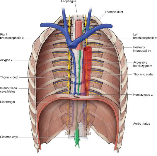
নিতু : বলছি, Posterior mediastinum is bounded anteriorly by pericardium and it’s contents, bifurcation of the trachea and pulmonary vessels, posteriorly by the bodies of lower eight thoracic vertebrae and intervening intervertebral discs, superiorly by transverse thoracic plane separating superior and inferior mediastinum, inferiorly by the diaphragm and on each side mediastinal pleura।
শিলা : ওকে, What are the contents?
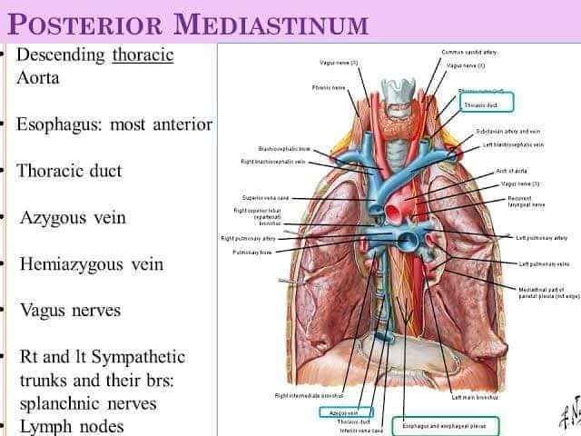
নিতু : Contents are esophagus, thoracic duct, descending thoracic aorta and it’s branches, azygos veins, hemiazygos veins, accessory hemiazygos veins, vagus nerves, sympathetic trunks and splanchnic nerves and posterior mediastinal lymph nodes।
শিলা : বল তো, What are the common structures that are found in both superior and posterior mediastinum?
নিতু : Esophagus and thoracic duct পাওয়া যায়।
শিলা : What is thoracic duct?
নিতু : The thoracic duct is the largest lymphatic vessel in the body, allowing return of lymph from most of the body into the venous system।
শিলা : গুড, বলতে পারবি body এর কোন side বা part থেকে thoracic duct drain করে না?
নিতু : Right superior quadrant, অর্থাৎ right side of head and neck, right upper limb, right lung and thoracic wall, right half of the heart and the convex surface of the liver।
শিলা : তাহলে right superior quadrant এর lymphatics কিসের মাধ্যমে drain হয়?
নিতু : Lymphatic duct এর মাধ্যমে।
শিলা : খুব ভালো। Thoracic duct lymphatics কোথায় drain করে?
নিতু : Drains into the angle of the left subclavian and internal jugular veins as a single trunk।
শিলা : হ্যাঁ, আচ্ছা বলে রাখি, common pathology of thoracic duct is chylothorax and lymphocele of the thoracic duct।
নিতু : আপু Chylothorax কি?

শিলা : A chylothorax is an accumulation of lymphatic fluid in the space surrounding the lung (pleural space) and it is caused by disruption of the thoracic duct and distributor resulting in chyli into pleural space।
নিতু : Chyli কি?
শিলা : lymphatics fluid of interstitial region। আচ্ছা বলতে পারবি, chyli দেখতে কেমন?
নিতু : না, আপু।
শিলা : Chyli এর রং সাদা, বলতে পারিস milky-white fluid।
নিতু : ওকে, আপু।
শিলা : এবার বল, What is esophagitis?
নিতু : এটা তো জানিনা আপু।
শিলা: Esophagitis means Inflammation of the esophagus এবং esophageal ulcer is an erosion in an area of the lining of the esophagus। আরো বিস্তারিত Esophagus পড়ার সময় জানতে পারবি।
নিতু : আপু, What are the causes of posterior mediastinal masses?
শিলা : Causes of posterior mediastinal masses include esophageal lesions, congenital or acquired vascular lesions, foregut cysts, intrathoracic goiters, mediastinal pseudocysts, fat-containing tumors, adenopathy, neurogenic tumors, infectious spondylitis, and vertebral tumors।
নিতু : Neurogenic tumors গুলো ও posterior mediastinum এর সাথে সম্পর্কিত?
শিলা : হ্যা, Up to 95% of neurogenic tumors occur in the posterior mediastinum, and they are the most common posterior mediastinal masses।
নিতু : আপু Thoracic aorta কিসের continuation ?
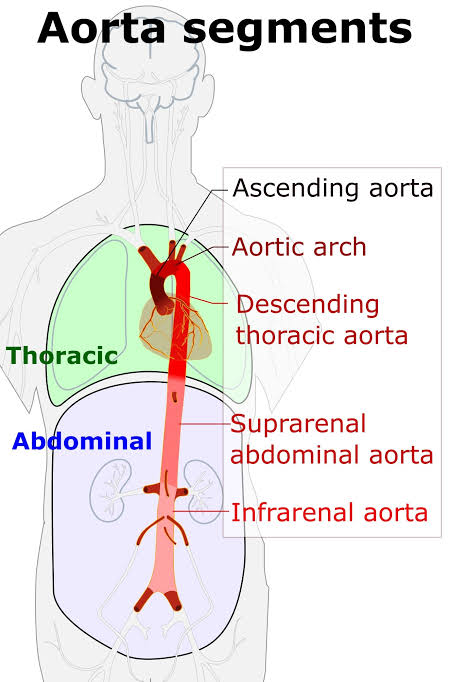
শিলা : Arch of aorta এর continuation, এবং posterior mediastinum এ thoracic aorta এর কিছু branches ও আছে, বলতে পারবি কি কি?
নিতু : হ্যাঁ, পারব, বলছি bronchial branches, pericardial branches, posterior intercostal branches, superior phrenic branches, esophageal branches and subcostal branches।
শিলা : এর bronchial branches গুলো trachea, bronchi and lymph nodes কে supply দেয়। আচ্ছা বল তো Azygos venous system কোথায় drain হয় ?
নিতু : It enters the mediastinum via aortic hiatus and drains into the superior vena cava।
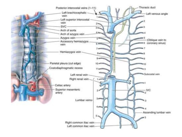
শিলা : হ্যাঁ, গুড, মূলত, The azygos system of veins consists of several longitudinal vessels, these are azygos, hemiazygos, accessory hemiazygos veins on both sides of the vertebral column that collect venous blood from the viscera of the mediastinum। Azygos venous system কিন্তু venous drainage এর জন্য খুবই গুরুত্বপূর্ণ, বিশেষভাবে lower part of the body এর জন্য।
নিতু : কেন আপু?
শিলা : কারণ, The azygos venous system is an essential anastomotic pathway that can return venous blood to the heart from the lower part of the body if the inferior vena cava is blocked।
নিতু : আচ্ছা আপু, posterior mediastinum এর সাথে neck এবং thigh এর কোনো clinical correlation আছে?
শিলা : হ্যাঁ আছে, খুব ভালো প্রশ্ন করেছিস, বলছি, মূলত, extension of pus into posterior mediastinum from neck এবং extension of pus into thigh from posterior mediastinum হতে পারে।
প্রথমত, pus from neck can extend into posterior mediastinum because of the spaces in the neck between pretracheal and prevertebral layers of deep cervical fascia such as retropharyngeal space।
এছাড়া pus from posterior mediastinum can easily enter the psoas sheath which communicates with the posterior mediastinum by a funnel shaped orifice and track down into the thigh in the region of femoral triangle।
নিতু : বুঝতে পেরেছি আপু।
শিলা : এবার বল Mediastinitis কি ?
নিতু : It is the inflammation of the loose connective tissue of the mediastinum।
শিলা : What is mediastinal shift?
নিতু : আপু এটা তো জানিনা।
শিলা : ওকে, মূলত the mediastinum shifts to the affected side due to appreciable reduction in lung volume, decrease in intrapleural pressure and as in collapse of lung and atelectasis।
Mediastinal shift can be detected by palpating the trachea in the suprasternal notch।
নিতু : ওকে আপু, আচ্ছা আপু widening of the mediastinum কি ?
শিলা : মূলত, when the mediastinum is greater than 6 to 8cm, depending on which source, it is noted to be wide।
The most significant life-threatening concern associated with a wide mediastinum is an acute aortic rupture।
নিতু : আপু , What are the causes of the widening of mediastinum?
শিলা : ওকে, mass or lymphadenopathy, cause focal widening, whereas acute causes of a widened mediastinum, like infection or bleeding, cause diffuse, generalized widening, etc।
নিতু : Pneumomediastinum কি আপু?
শিলা : মূলত pneumomediastinum হলো presence of air in the mediastinum, which in some cases can lead to pneumothorax, pneumoperitoneum, and pneumopericardium if left untreated
নিতু : আপু সত্যিই অনেক কিছু জানতে পারলাম, ধন্যবাদ আপু 😇।
Reference :
🟢 Gray’s Anatomy for Students 4th Edition 🟢 A. K. Datta, Essentials of Human Anatomy, ( 10th edition )
🟢 Vishram Singh, Textbook of Anatomy, Volume – 1( 2nd edition )
Platform Academic Division/
Ohia Farzin Raha,
CARe Medical College,
session : 2019-20.