
আজ meningeal haemorrhage নিয়ে আলোচনা করছি।
Brain কে কভার করে থাকে ৩ স্তর বিশিষ্ট meninges। বাইরে থেকে ভেতরে, স্তরগুলো হলোঃ
- Dura mater
- Arachnoid mater
- Pia mater
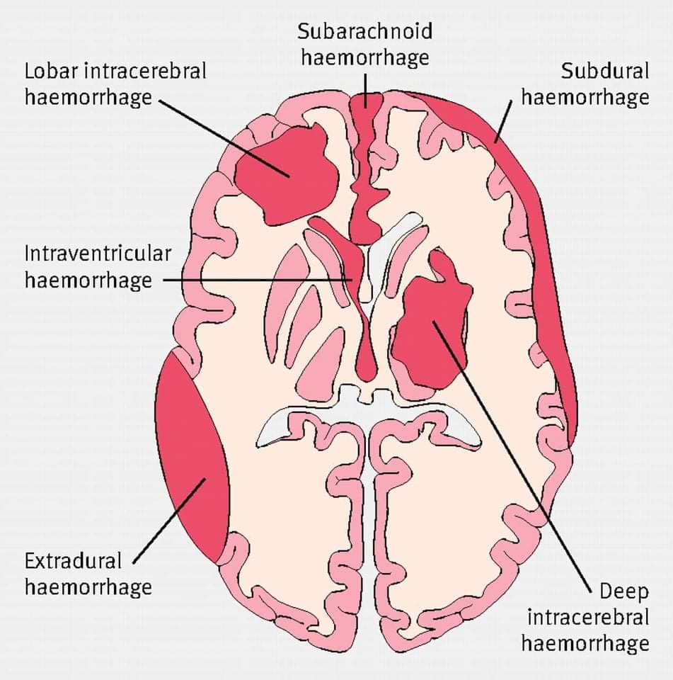
যেকোনো দুটি স্তরের মধ্যবর্তী জায়গায় কোনো কারণে রক্তক্ষরণ হলে সেটাকে বলে meningeal haemorrhage/haematoma।
Meningeal haemorrhage ৪ প্রকার, যা নিচে একে একে আলোচনা করা হয়েছে।
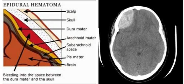
Epidural haematoma:
It is the collection of blood in the potential space between the skull and periosteal layer of the dura mater.
Epidural haematoma র সব থেকে কমন কারণ হচ্ছে fracture of the temporal bone.
Temporal bone এর একটি অত্যন্ত পাতলা অংশ হচ্ছে pterion। এই pterion এ খুব সহজেই fracture হয়ে যেতে পারে।এর ফলে, middle meningeal artery র laceration হয়ে bleeding শুরু হয়ে যায়, যা epidural space এ accumulate হওয়ার কারণে haematoma র সৃষ্টি হয়।
🚩Symptoms:
- সব থেকে গুরুত্বপূর্ণ উপসর্গ হলো lucid interval। (Lucid interval is defined as the brief period of consciousness between two unconscious periods.)
- Headache
- Nausea
- Vomiting
- Focal neuro-symptoms
- 3rd cranial nerve palsy
🚩Diagnosis এর উপায় কী?
এক কথায় CT-scan.
Epidural haematoma suture line ক্রস করতে পারে না। তাই CT scan এ biconvex / lens-shaped haematoma দেখা যাবে।
🚩 Treatment :
- Evacuate the haematoma by: Craniotomy or Burr hole surgery .
- Maintenance of intra-cranial pressure
এরপর কথা বলব subdural haematoma নিয়ে।
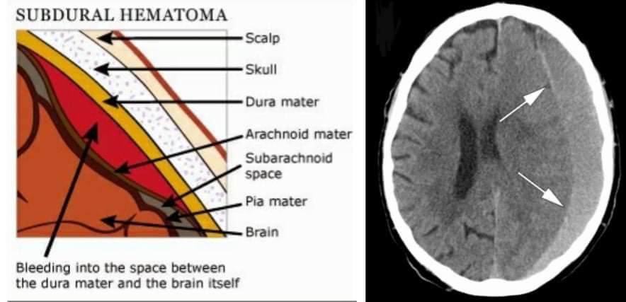
Subdural haematoma:
নাম শুনেই বোঝা যাচ্ছে এই haematoma টি subdural space, অর্থাৎ meningeal layer of dura mater আার arachnoid mater এর মধ্যবর্তী জায়গাটিতে হয়।
🚩Subdural haematoma কেন এবং কীভাবে হয়?
Brain এর সাথে epidural space এ অবস্থিত dural venous sinus এর সাথে সংযোগ স্থাপনকারী vein গুলোর নাম হচ্ছে bridging vein।
এই bridging vein যদি কোনো কারণে ছিঁড়ে যায়, তখন subdural haematoma র সৃষ্টি হয়।
🚩Subdural haematoma সাধারণত কোন কোন ক্ষেত্রে হয়ে থাকে?
Motor vehicle accident, trauma, fall কিংবা বয়স্ক ব্যক্তিদের subdural haematoma হতে পারে।
🚩Symptoms:
- Headach
- Nausea
- Vomiting
- High blood pressure
- Low pulse
- Confusion
🚩Diagnosis :
CT scan করতে হবে।
রিপোর্টে crescent-shaped/ concave haematoma দেখা যাবে। উল্লেখ্য এই ধরনের haematoma, suture line ক্রস করতে সক্ষম।
🚩Treatment :
Evacuate the haematoma by:
- Craniotomy
- Burr hole surgery
এরপর subarachnoid haemorrhage।
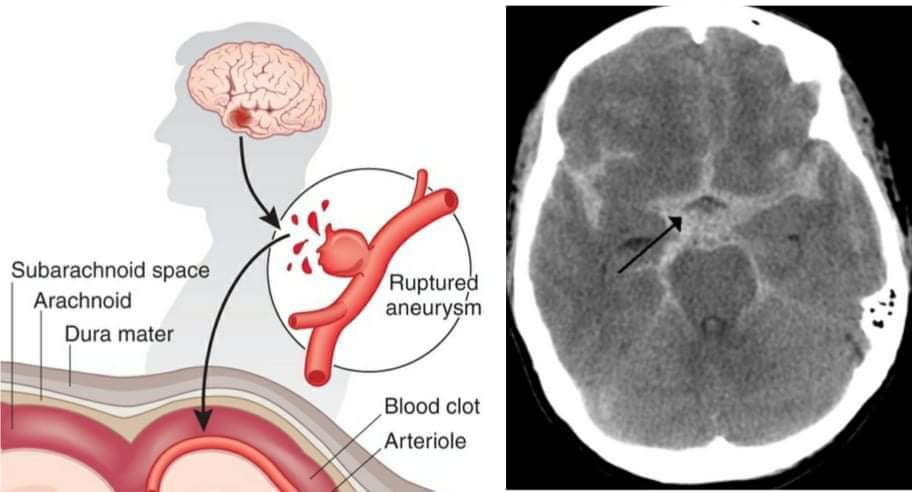
Subarchnoid haemorrhage:
It is the bleeding which occurs in the subarachnoid space due to rupture of any aneurysm.
বেশিরভাগ সময় anteriror communicatng artery অথবা middle cerebral artery র Berry/Saccular aneurysm rupture এর কারণে subarachnoid haemorrhage হয়।
🚩Symptoms:
- Thunderclap headache। এই উপসর্গটিই মূলত subarachnoid haemorrhage কে বাকিগুলো থেকে আলাদা করে।
- Nausea
- Vomiting
- Meningeal symptoms (i.e. neck stifness, photophobia, delirium)
🚩Diagnosis:
যথারীতি CTscan করতে হবে।
- যদি রিপোর্ট পজিটিভ আসে, তাহলে 4 vessel angiography ও করতে হবে।
- নেগেটিভ আসলে lumbar puncture করতে হবে।
- Lumbar puncture এর মাধ্যমে যেই CSF সংগ্রহ করা হবে, তাতে xanthochromia থাকবে।
🚩Treatment:
•Endovascular coiling/ clipping
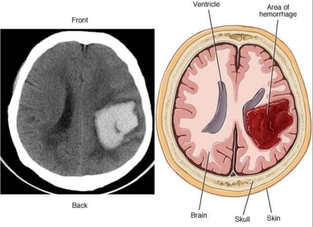
লাস্ট বাট নট দা লিস্ট, Intracerebral haemorrhage
Intracerebral haemorrhage: It is the bleeding which occurs within the brain substance.
সাধারণত thalamus, basal ganglia, cerebellum এবং pons এ bleeding হয়ে থাকে।
🚩 Cause of bleeding:
- Hypertension
- Arterio-venous malformation
- Leptomeningeal amyloidosis
🚩Symptoms:
- Headache
- Nausea
- Vomiting
- Hemiparesis
- Hemiplegia
- Loss of consciousness
🚩 Diagnosis:
- ঠিক ধরেছেন CT scan.
- *** Intra-cerebral bleeding থাকলে কখনই lumbar puncture করা যাবে না!!!!
🚩 Treatment:
- Evacuate the haematoma
- Give mannitol to reduce intra-cranial pressure.
Fahima Hasan
MH Samorita Medical College
2017-18