
Nail Abnormality and Associated systemic disease:
Let’s study them all in one post!!
★ Clubbing:
Definition: Clubbing (also called Hippocratic fingers) is defined as a selective bulbous enlargement of the distal segment of a digit (fingers and toes) due to an increase in connective tissue especially on the dorsal aspect resulting in loss of the onychonychia angle.
Causes of clubbing:
A) Respiratory:
- Neoplasms: Bronchogenic carcinoma (especially squamous cell carcinoma), metastasis to lung, mesothelioma.
- Suppurative lung disease: Bronchiectasis, lung abscess, cystic fibrosis, empyema.
- Asbestosis (with mesothelioma).
- Sarcoidosis.
- Cystic fibrosis.
- Pulmonary arteriovenous fistula.
- Interstitial lung diseases.
B) Cardiovascular:
- Infective endocarditis.
- Cyanotic congenital heart diseases.
- Atrial myxoma.
- Eisenmenger’s syndrome.
C) Gastrointestinal:
- Inflammatory bowel disease (Ulcerative colitis and Crohn’s disease).
- Primary biliary cirrhosis.
- Hepatoma.
D) Miscellaneous:
- Hereditary, idiopathic: (Pachydermoperiostosis or Touraine-Solente-Gole syndrome).
- Unidigital clubbing occurs in repeated trauma.
- Unilateral Clubbing occurs in Hemiplegia (long standing).
- Vascular disease:
– Aneurysm: Subclavian artery, brachiocephalic trunk.
– Pre-subclavian coarctation of aorta (left-sided clubbing).
– Pancoast tumor.
– Unilateral erythromelalgia.
– Arteriovenous fistula used for hemodialysis.
– Infected arterial graft.
★ Koilonychia:
Spoon-shaped nails (transverse and longitudinal concavity)
Found in:
a. Iron deficiency anemia,
b. Rarely in hemochromatosis,
c. Raynaud’s disease,
d. Systemic lupus erythematosus,
e. Hypothyroidism or hyperthyroidism.
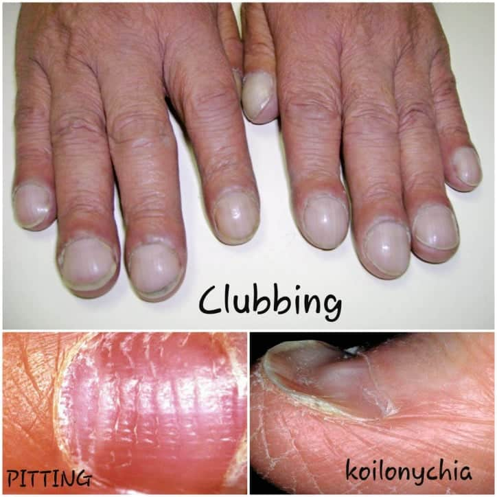
★ Pitting
Punctate depressions in nails.
Found in:
a. Psoriasis,
b. Reiter’s syndrome,
c. Pemphigus,
d. Lichen planus,
e. Alopecia areata,
f. Rheumatoid arthritis.
★ Onycholysis:
Distal nail plate separated from nail bed, white discoloration of the affected part of the nail.
Found in:
a. Psoriasis,
b. Local infection,
c. Hyperthyroidism,
d. Sarcoidosis,
e. Trauma,
f. Amyloidosis,
g. Connective tissue disorders,
h. Pellagra.
★ Beau’s lines:
Transverse linear depressions over nails, move distally with the growth of nail.
Found in:
a. Any severe systemic illness that disrupts nail growth,
b. Raynaud’s disease,
c. Pemphigus,
d. Trauma.
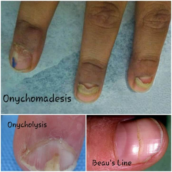
★ Onychomadesis:
Proximal separation of nail plate from nail bed.
Found in:
a. Trauma,
b. Drug sensitivity,
c. Poor nutrition,
d. Pemphigus vulgaris,
e. Kawasaki disease.
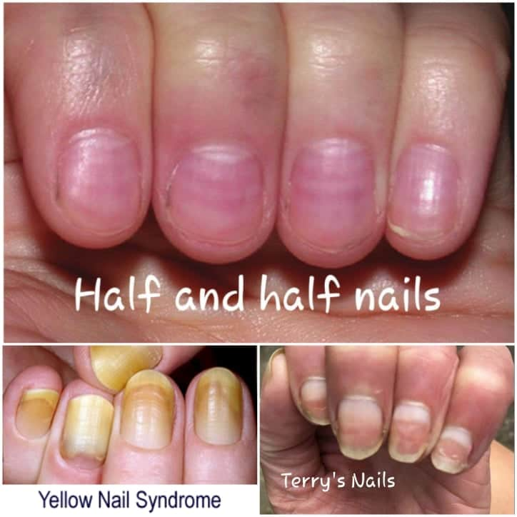
★ Yellow nails:
Nail has a ‘heaped-up’ or thickened appearance, yellow in color, with obliteration of lunula.
Found in:
a. Lymphedema,
b. Pleural effusion,
c. Immunodeficiency,
d. Bronchiectasis,
e. Sinusitis,
f. Rheumatoid arthritis,
g. Nephrotic syndrome,
h. Thyroiditis,
i. Tuberculosis,
j. Raynaud’s disease.
COLOR CHANGE
★ Terry’s (white) nails:
Most of the nail plate turns white with obliteration of lunula, uniformly affects all nails.
Found in:
a. Liver failure,
b. Cirrhosis,
c. Diabetes mellitus,
d. Congestive heart failure,
e. Hyperthyroidism,
f. Malnutrition.
★ Half and half nails (Lindsay’s nails):
Proximal portion of nail bed is white because of nail bed edema (half brown, half white appearance).
Found in:
a. Renal failure,
b. HIV infection,
c. Crohn’s disease.
★ Azure lunula (blue nails):
Found in:
a. Hepatolenticular degeneration (Wilson’s disease),
b. Silver poisoning,
c. Quinacrine therapy.
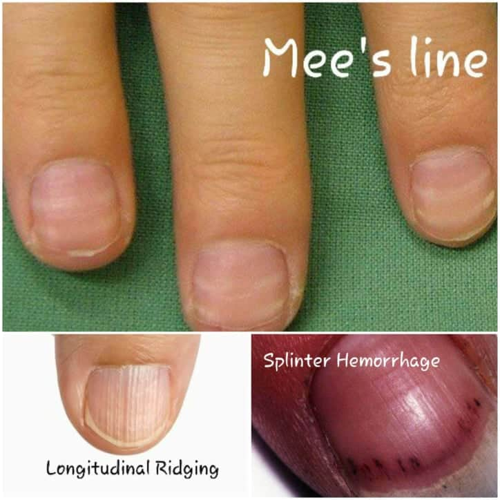
★ Mees’ lines:
Transverse white bands affecting multiple nails, move distally with nail growth.
Found in:
a. Arsenic poisoning,
b. Hodgkin’s lymphoma,
c. Congestive heart failure,
d. Leprosy,
e. Malaria,
f. Chemotherapy.
★ Muehrcke’s lines:
Pairs of transverse white lines that disappear on applying pressure, do not move with growth of nail.
Found in:
a. Specific for hypoalbuminemia of any cause.
★ Dark longitudinal streaks:
Found in:
a. Melanoma,
b. Benign nevus,
c. Chemical staining,
d. Normal variant in darkly pigmented people.
★ Splinter hemorrhage:
Longitudinal, thin, reddish lines occurring beneath the nail plate.
Found in:
a. Subacute bacterial endocarditis,
b. Systemic lupus erythematosus,
c. Rheumatoid arthritis,
d. Peptic ulcer disease,
e. Malignancies,
f. Oral contraceptive use,
g. Pregnancy,
h. Psoriasis,
i. Trauma.
★ Telangiectasia:
Irregular, twisted and dilated vessels at the distal portion of cuticle covering the nail bed.
Found in:
a. Rheumatoid arthritis,
b. Systemic lupus erythematosus,
c. Dermatomyositis,
d. Scleroderma.
★ Longitudinal striations:
Found in:
a. Alopecia areata,
b. Vitiligo,
c. Atopic dermatitis,
d. Psoriasis.
Ashraful Emdad,
Army Medical College Cumilla,
Session: 2016-17.
Platform academic / Ariful Islam Neloy