
The anterior compartment of the thigh is one of the three compartments in the thigh. Muscles within this compartment primarily produce hip flexion and knee extension. The thigh is separated into anterior, posterior and medial (adductor) compartments by intermuscular septa and surrounded by the fascia lata.
★ Contents:
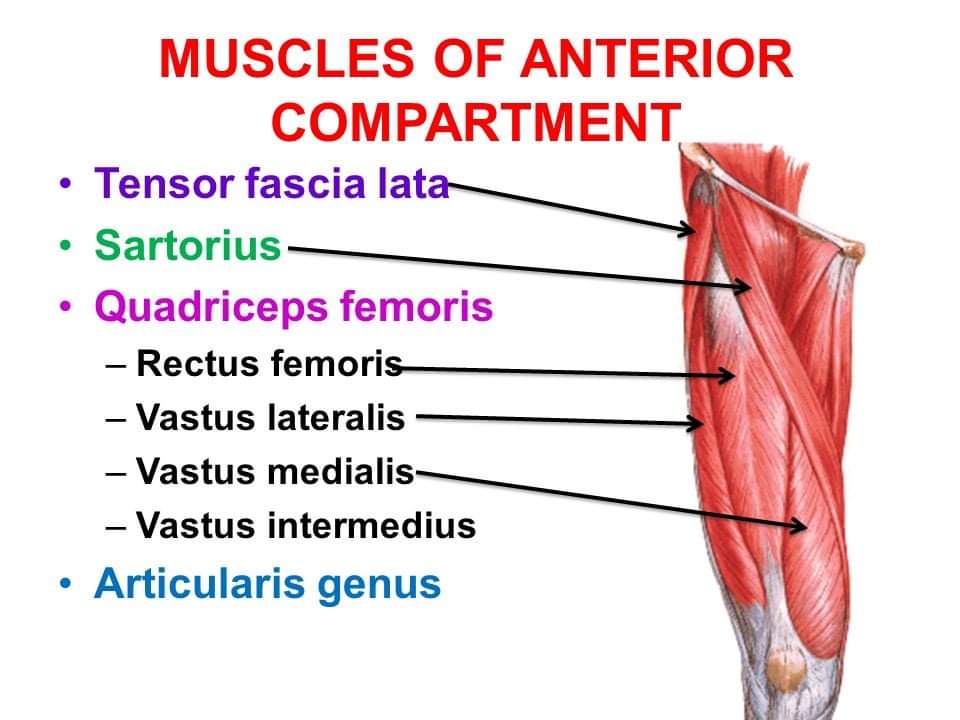
◾ Muscles: Quadriceps femoris, articularis genu, sartorius and tensor fascia latae.
◾ Nerve: Femoral nerve.
◾ Artery: Femoral artery.
★ Origin, insertion and actions of the front of the thigh:
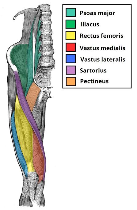
⬛ Quadriceps femoris: This muscle is so named because it consists of four parts: rectus femoris, vastus lateralis, vastus medialis and vastus intermedius.
(A) Rectus femoris:
🔸 Origin: Straight head from the upper half of the anterior inferior iliac spine. Reflected head from the groove above the acetabulum.
🔸 Insertion: Base of patella.
🔸 Actions: Flexion of the hip joint and extension of the knee joint.
(B) Vastus lateralis:
🔸 Origin: Upper part of the intertrochanteric line; Anterior and inferior borders of greater trochanter.
🔸 Insertion: Base and upper 1/3rd of the lateral border of the patella.
🔸 Action: Extension of the knee joint.
(C) Vastus medialis:
🔸 Origin: Lower part of the intertrochanteric line; Spiral line; Medial lip of the linea aspera.
🔸 Insertion: Base and upper 2/3rd of the medial border of the patella.
🔸 Action: Extension of the knee joint.
(D) Vastus intermedius:
🔸 Origin: Lateral surface of the shaft of the femur.
🔸 Insertion: Base of the patella.
🔸 Action: Extension of the knee joint.
⬛ Articularis genu
🔸 Origin: Anterior surface of the lower part of the shaft of femur.
🔸 Insertion: Synovial membrane of the knee joint.
🔸 Action: Pulls up the synovial membrane of the knee joint during it’s extension.
⬛ Sartorius
🔸 Origin: Anterior superior iliac spine and upper half of the notch below it.
🔸 Insertion: Upper part of the medial surface of the shaft of the tibia.
🔸 Action: Flexion of the hip and knee joints; Lateral rotation of the thigh.
⬛ Tensor fasciae latae
🔸 Origin: Outer lip of the iliac crest from anterior superior iliac spine to the tubercle of iliac crest.
🔸 Insertion: Iliotibial tract 3-5 cm below the greater trochanter.
🔸 Action: Abduction of the hip joint.
🔵 Femoral triangle: It is a triangular depression on the front of the upper 1/3rd of the thigh below the inguinal ligament.
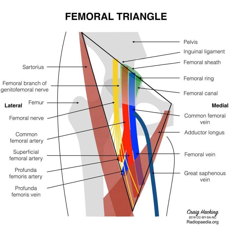
🔷 Boundaries of femoral triangle:
✔️ Laterally: By the medial border of sartorius.
✔️ Medially: By the medial border of adductor longus.
✔️ Base: Formed by inguinal ligament.
✔️ Apex: By meeting point of the sartorius and adductor longus.
✔️ Floor: By iliacus, psoas major, pectineus and adductor longus.
✔️ Roof: By skin and fascia.
🔷 Contents:
✔️ Femoral artery and it’s branches.
✔️ Femoral vein and it’s tributaries.
✔️ Femoral nerve.
✔️ Deep inguinal lymph nodes.
✔️ Lateral cutaneous nerve of the thigh.
✔️ Femoral branch of genitofemoral nerve.
✔️ Fibrofatty tissue.
🔵 Femoral sheath: It is a funnel shaped fascial sheath enclosing the upper 3-4 cm of the femoral vessels.
🔴 Adductor canal/ Hunter’s canal: The adductor canal is a long intermascular tunnel situated on the medial side of the middle 1/3rd of the thigh.
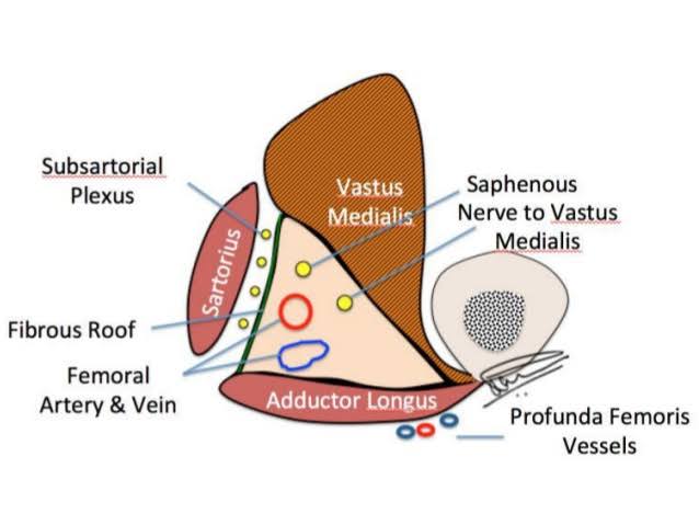
🔷 Contents:
✔️ Femoral artery.
✔️ Femoral vein.
✔️ Saphenous nerve
✔️ Nerve to vastus medialis.
✔️ Anterior and posterior divisons of the obturator nerve.
✔️ Descending genicular artery, a branch of the femoral artery.
🔷 Clinical significance of adductor canal: The femoral artery is exposed and ligated in the adductor canal during surgery for aneurysm of the popliteal artery.
★ Femoral nerve: It is the cheif nerve of the anterior compartment of the thigh. It is the largest branch of the lumbar plexus and arises from the dorsal divisons of the anterior primary rami of L1, L2, L3 nerves.
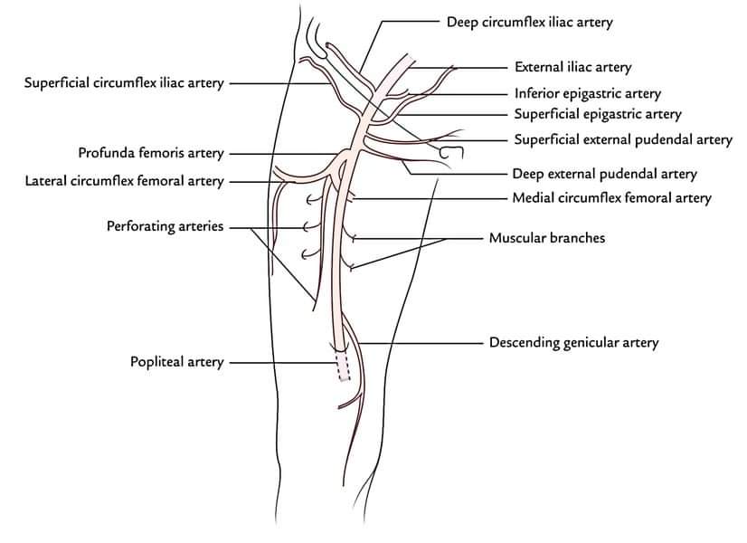
🔴 Femoral artery: It is the continuation of external iliac artery and enters the femoral triangle behind the inguinal ligament at the midinguinal point.
🟥 Branches: In the femoral triangle:
◾ Three superficial branches: Superficial epigastric artery, superficial external pudendal artery and superficial circumflex iliac artery.
◾ Three deep branches: Profunda femoris artery, deep external pudendal artery and muscular branches.
🔵 Femoral vein: It is the upward continuation of the popliteal vein at the adductor hiatus.
🔷 Tributaries:
🔹 Great saphenous vein.
🔹 Profunda femoris artery.
🔹Medial and lateral circumflex femoral veins.
🔹 Deep external pudendal vein.
🔹 Direct mascular tributaries.
🔺 Reference: Textbook of anatomy by Vishram Singh( Volume 2).
Platform academic divison/ Mehedi Hasan Shariar.
Central medical college, Cumilla.
Session: 2019-2020.